in Foot and Ankle Surgery and Reconstruction
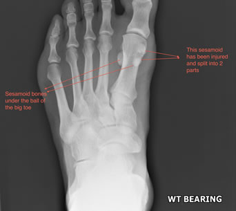
A sesamoid bone is a bone that lies within a tendon. The big example is the patella or kneecap bone lying mobile in the front of the knee within the thigh muscle tendon. In the foot the “SESAMOIDS’’ refer to 2 tiny bones under the ball of your big toe. Each one is about 1x1.5cm in size and helps the big toe joint moving from a straight position to a bent or flexed position.

Different problems can arise at the sesamoid bones:
The clinical story can indicate whether an acute injury is likely or a more persistent chronic problem exists. An X-Ray can determine whether the bone is positioned correctly or even fractured. Special X-Ray views can be taken to look accurately at the sesamoid bones under the big toe. Other special tests can be helpful including an MRI scan or a CT scan, but your surgeon will advise on this as required.
If the symptoms are associated with an acute injury, rapid surgical assessment is advised to ensure return to full function is assured. In the presence of a chronic, grumbling pain in the toe it is likely that it will continue to persist without surgical assessment, where a variety of treatments may be applicable.
In the presence of an acute injury please refer to turf toe injuries
For more chronic patterns of symptoms:
Surgical intervention can follow other non-invasive therapies if unsuccessful. The sesamoid bones can be removed if troublesome however the surgeon must ensure correct reattachment of important tendons that were attached to the sesamoids to ensure good toe strength. If the sesamoids have been fractured then fixation with a screw can be done. Return to a good level of activity should be possible following surgery.
Many patients are simply seeking advice on managing a problem. If non-operative measures have been unsuccessful and your quality of life is affected then surgery can always be discussed with your surgeon who will inform you of the potential outcomes from the surgical intervention planned.